What is a Dysplastic Nevus? By Jacob Anderson, B.S, Anais Hacobian, B.S and Dr. David Robles MD, PhD
What is a Dysplastic Nevus?
Dysplastic nevi are classified as moles that can occur anywhere on the body; but they differ in size, shape and color when compared to the common mole. Compared to the common mole, dysplastic nevi are typically, but not always, larger than 5 millimeters in size, unevenly colored with a wide range of colors that includes pink to dark or light brown.


Demographics and Causes?
Dysplastic nevi can occur in any individual and at any age. They are most typically caused by ultraviolet radiation (UV) and genetics.
UV Radiation
Atypical moles are often found on areas of the skin that are exposed to ultraviolet radiation (sunlight). Although dysplastic nevi can occur on all parts of the body, even those not exposed to sunlight, parts of the body constantly exposed to sunlight have increased chances of developing dysplastic nevi. Dysplastic nevi are considered precancerous moles, which is why UV exposure can increase the chance of developing melanoma.
Genetics (personal and family history)
An individual's personal and family history plays a role in developing dysplastic nevi. If an individual has a personal history of melanoma, they have an increased risk of developing dysplastic nevi as well as redeveloping melanoma. A family history of atypical moles and melanoma can also increase the risk of personally developing dysplastic nevi and/or melanoma.


How do you treat Dysplastic nevi?
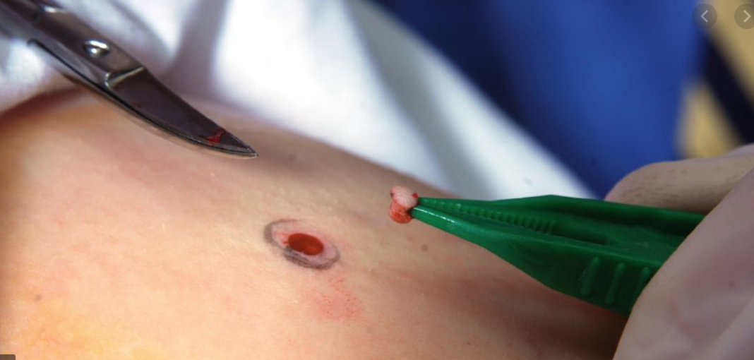
When considering treatment, Dermatologists consider these three most common options: biopsy, leave it alone and monitor, and excision. When dermatologists find irregularities, they often take a biopsy of the mole(s) to rule out any potential malignancy.
 |
| The above image shows the difference between the shave, punch or excisional biopsy |


If dysplasia (abnormal growth) is present, discussion about specific treatments is discussed. If there is only mild dysplasia, patients may just opt for monitoring the lesion for changes. Those with moderate to severe dysplasia are often excised. Newer data suggests that close observation is a reasonable approach for moderately dysplastic nevi.
What can I do for prevention?
Since dysplastic nevi are commonly seen in areas with the most sun exposure, the first component to take into consideration is being proactive in applying or wearing sun protection, and avoid tanning. For individuals who already have dysplastic nevi, limiting sun exposure will reduce the possibility of the development of cancer.
Patients can also monitor the nevus by using an acronym that Dermatologists often use when evaluating moles; become familiar with the ABCDE’s: A= asymmetry, B=border, C= color, D= diameter and E= evolving. If your mole(s) consists of non-symmetrical, non-uniform borders and edges, color changes, growing in size and changing in appearance, this is a good indication to contact your Dermatologist for further assessment and a skin check.

Comments
Post a Comment